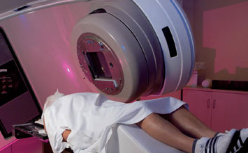
Minimal invasive surgery (MIS) is aiming at causing patients as little discomfort as possible and endoscopes play an important role here. Traditional Chip-on-the-Tip endoscopes use fixed focus optics, which cannot display objects at different distances with optimum resolution. By integrating a miniature drive, it could become possible to achieve variable focusing, so that the object can always be displayed optimally in sharp focus.
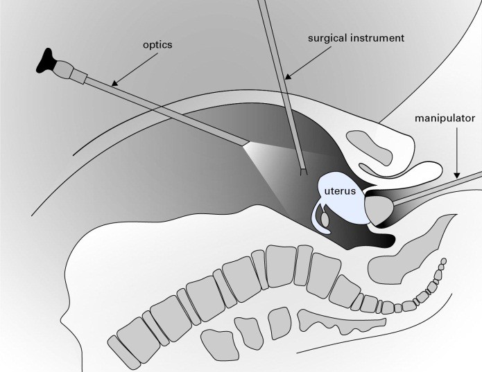
Endoscopes that allow minimally invasive surgery (MIS) are used in larynx diagnostics or laparoscopy (a surgery that uses a thin tube fed through an incision in the belly to look at the abdominal organs). Optics and special instruments (scissors, hooks, grasping forceps, ultrasound scissors, etc.) are then introduced into the abdominal cavity through so-called working trocars. Following surgery, the patient experiences less post-operative pain and can be discharged from the hospital earlier, due to the quicker healing process. The risk of wound infections or wound healing disorders is minimized and allows treatment of high-risk patients.
Placing the Imaging Sensor on the Endoscope Tip
Traditional endoscope technology is well established and has reached a high degree of sophistication. One of the bottlenecks used to be the image transmission through classical optics. With the rapid evolution of microelectronics and compact imaging sensors, more progress is feasible, such as integrating a small camera into the endoscope tip (Chip-on-the-Tip). The advantage is vastly improved image quality.
With traditional endoscope optics, it was, however, possible to attach a manual zoom lens or focus lens between the eyepiece and camera to set the focus to different object distances and zoom into details. In contrast, the digital zoom function of the Chip-on-the-Tip technology only allows a detail to be enlarged, which is always accompanied by a loss in quality. The absence of a focus function requires a compromise between depth of focus and image brightness.
Optical Zoom Function for Chip-on-the-Tip Cameras in Endoscopes
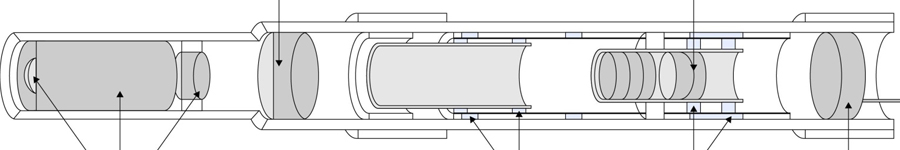
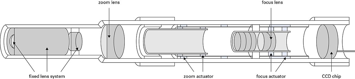
This type of compromise could soon be a thing of the past, if the actuators required for optical focus and zoom functions could be installed between the optics and the imaging chip. With diameters of typically 10mm, the potential installation space is very small and it seems to be difficult, but not impossible, to find the right drives for the zoom and focus lens.
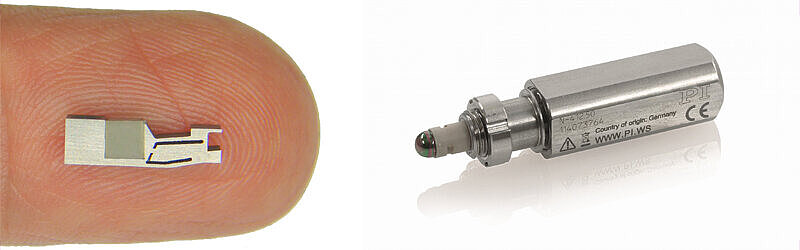
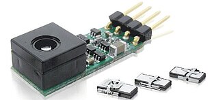
For example, miniaturized ceramic piezo motors and voice coil drives could open up a new area of application in this field. This is where PI (Physik Instrumente) and its leadership in miniature precision drive mechanisms come into play. These drives offer a solid base for use in modern high-resolution microscopes.
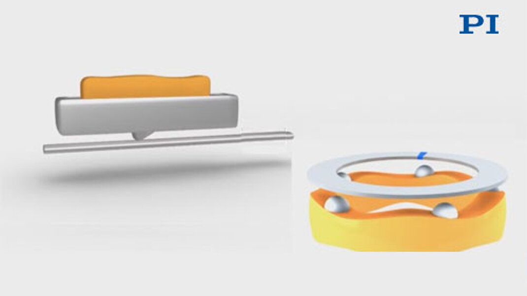
Small Piezo Motors or Voice Coil Drives
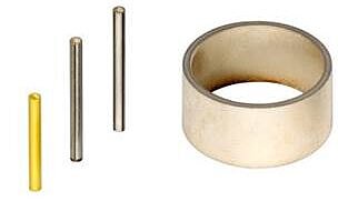
Piezo motors make for very compact and precise linear drives. The direct-drive principle eliminates the conversion of rotation into linear motion, along with the need for gears and other mechanical components that can wear and limit reliability. Piezo motors are small, lightweight, and consume very little energy. In fact, they can hold a position without power consumption.
There are a number of different piezo motor principles (see videos belows) that are feasible. All provide the necessary precision. Ultrasonic motors and inertia motors are very compact and cost effective.
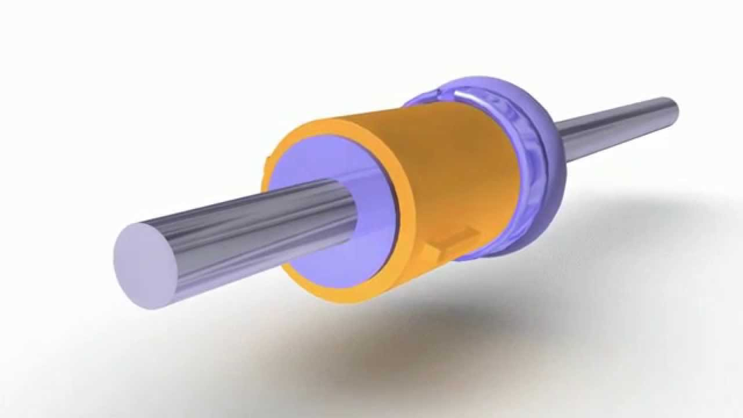
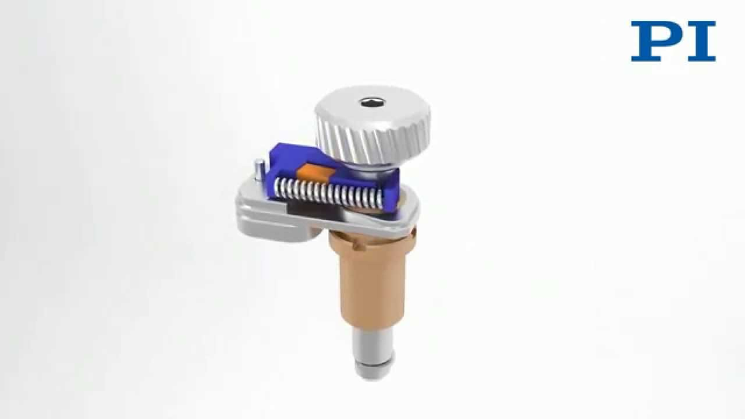
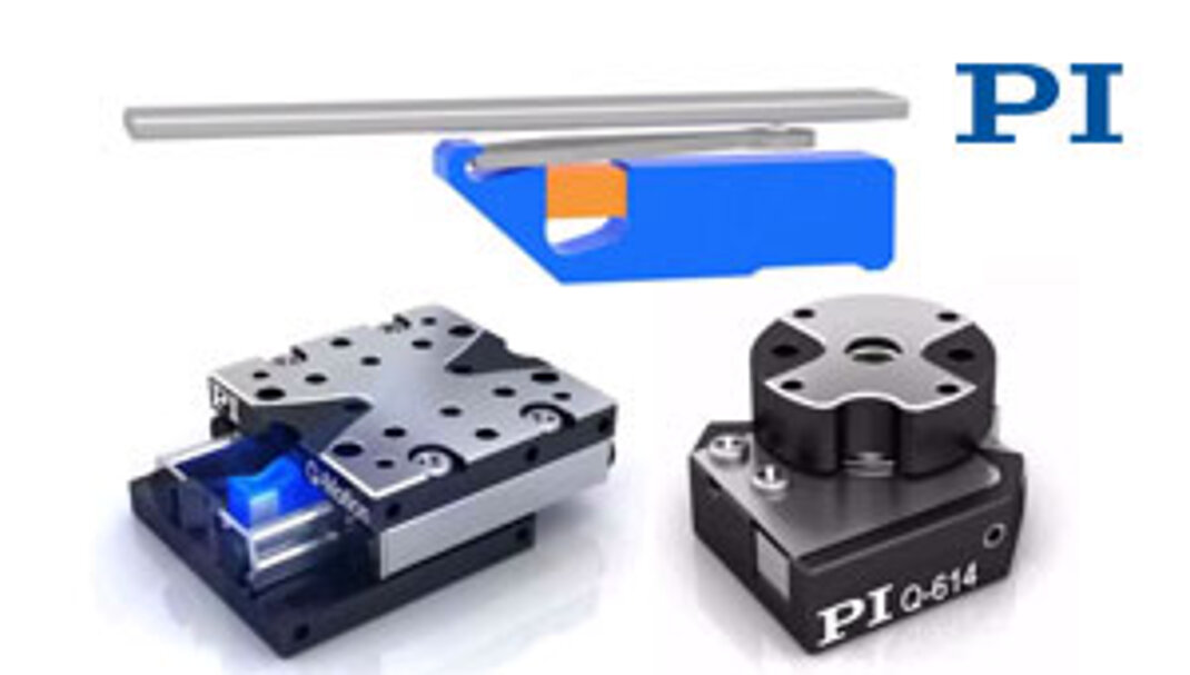
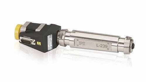
In addition to piezoelectric motors, traditional electromagnetic linear motors (Voice Coils) are also an option.
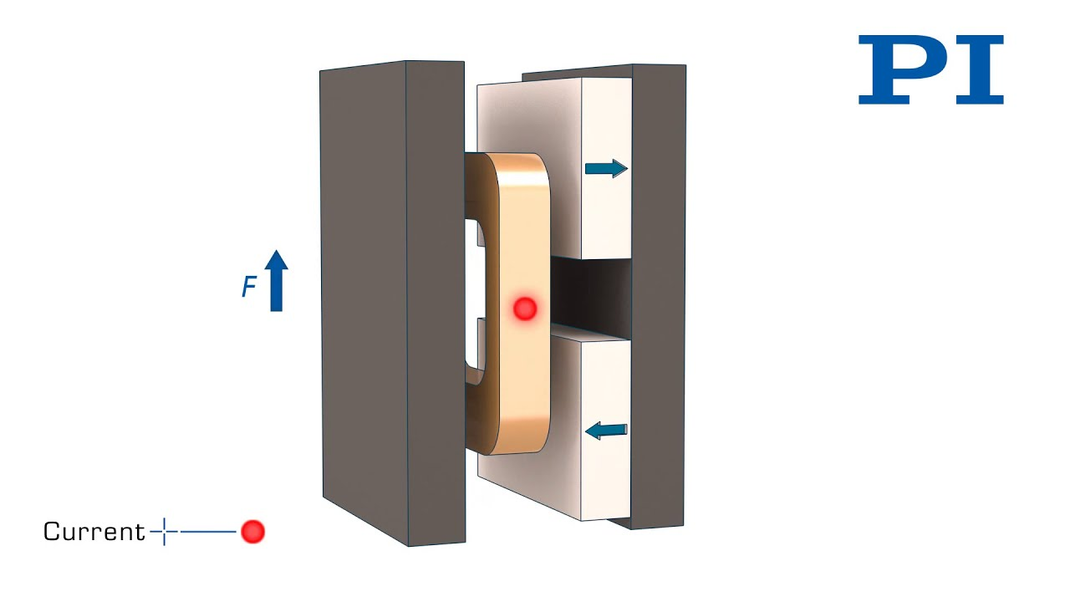
Voice coil actuators are related to the transducers used in loudspeakers. They can be manufactured with very small dimensions and provide motion ranges in the millimeter to centimeter range, just enough for endoscopy. Given these drive options, it should be interesting which of them will contribute to better image quality and depth of focus in the next generation of Chip-on-the-Tip endoscopes.
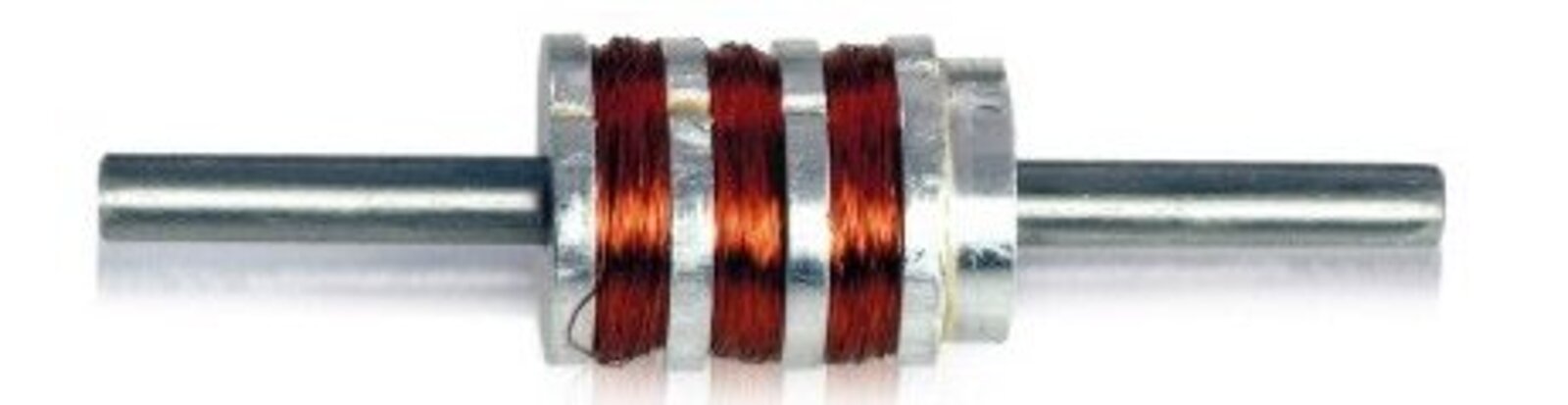
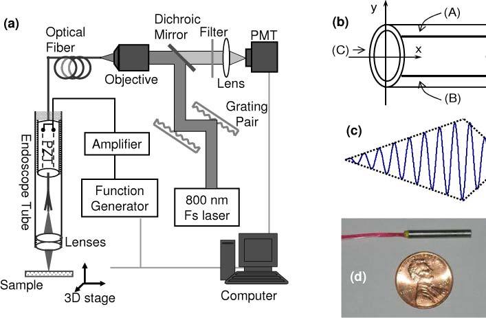
Piezo Scanner Tubes for 2-Photon Endoscopy
Compact piezo XY scanners can be used for high-resolution two-photon endoscopy. A recent paper describes a spiral fiber scanner based on a miniature piezo scanner tube (1.5mm outer diameter) used for biological imaging of parts of the eye. The compact instrument has a 2mm diameter providing lateral and axial resolution as low as 1.5μm.
Author: Birgit Bauer, Business Development Manager Health Care at PI (Physik Instrumente)


Blog Categories
- Aero-Space
- Air Bearing Stages, Components, Systems
- Astronomy
- Automation, Nano-Automation
- Beamline Instrumentation
- Bio-Medical
- Hexapods
- Imaging & Microscopy
- Laser Machining, Processing
- Linear Actuators
- Linear Motor, Positioning System
- Metrology
- Microscopy
- Motorized Precision Positioners
- Multi-Axis Motion
- Nanopositioning
- Photonics
- Piezo Actuators, Motors
- Piezo Mechanics
- Piezo Transducers / Sensors
- Precision Machining
- Semicon
- Software Tools
- UHV Positioning Stage
- Voice Coil Linear Actuator
- X-Ray Spectroscopy