Nondestructive Analysis of Tiniest Samples
At the Beamline U41-TXM of the electron storage ring BESSY II, a novel full-field transmission X-ray microscope (TXM) for the soft X-ray range was set up for the characterization of the nanostructure, chemical nature, and composition of materials on a 10 nm scale. Additionally to nano-tomography, a fluorescence light microscope has been developed that allows non-destructive analysis of tiniest samples of some micrometers, such as the distribution of heavy metals in anorganic or historic samples, and find answers for life sciences, geology, and material science.

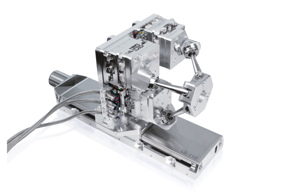
Positioning the Condenser Optics
The condenser optics of the X‑ray microscope is a lead crystal capillary that focuses the high‑energy synchrotron beam of about 300 µm diameter. The highly focused excitation radiation allows for analysis in the ppm range of a nanogram probe; elements of femtograms can be traced.
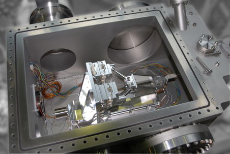
This capillary is positioned using a 7‑axis positioning system consisting of a long‑range linear stage with a 6‑axis SpaceFAB on top. The synchrotron beam passes through the aperture to the capillary condenser optics. Due to the high‑vacuum environment of 10‑7 hPa, all axes are equipped with vacuum‑compatible stepper motors.
Excellent position repeatability and high stiffness were essential to achieve the required accuracy. The SpaceFAB’s parallel-kinematics design allows for arbitrary setting of the pivot point by software commands. Thus, the operation can be adapted for different focusing optics easily.
Incorporated Fluorescence Light Microscope
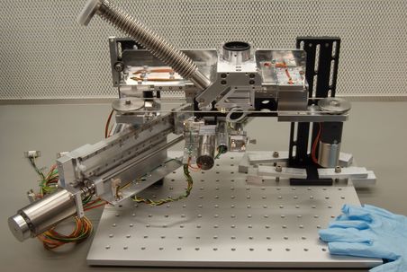
Integrated in the same vacuum chamber is a setup for fluorescence light detection. Both analytical methods can thus be performed on the same sample under identical conditions, allowing new insights on the structure.
This setup was commissioned as an addition to the already existing microscope, and thus had to follow the extra limitation to fit into the vacuum chamber. PI miCos designed and delivered the full setup ready to fit into the vacuum chamber, from light funnel to objective positioning to filter change system.